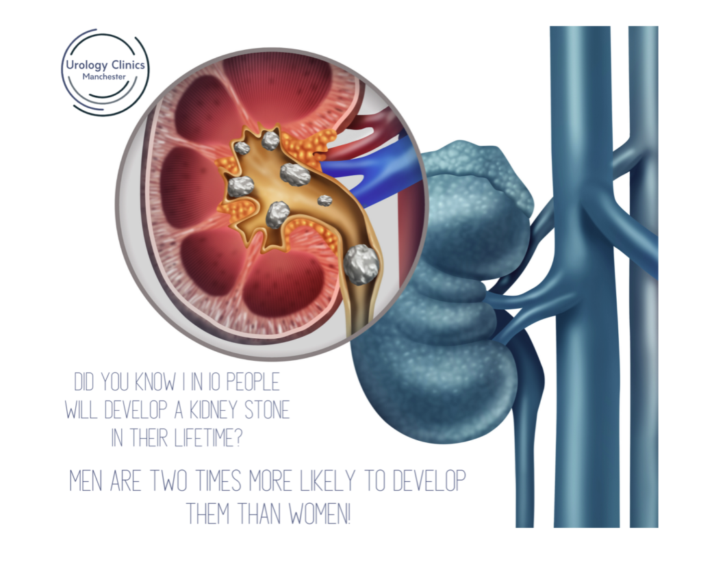
A distal ureteric stone, also known as a distal ureteral stone, refers to a kidney stone that has moved from the kidney and is located in the lower part of the ureter—the tube that connects the kidney to the bladder. Kidney stones are solid mineral and salt deposits that can form in the kidneys and may vary in size.
Here are some key points related to distal ureteric stones:
Location:
The term “distal” indicates that the stone is located in the lower part of the ureter, closer to the bladder. Stones in this location can cause symptoms and may affect urine flow.
Symptoms:
Distal ureteric stones can cause symptoms such as severe pain, typically on one side of the lower back or abdomen. This pain is often referred to as renal colic and can radiate to the groin and genital area. Other symptoms may include frequent urination, urgency to urinate, and blood in the urine.
Obstruction:
When a stone is in the distal ureter, it may obstruct the flow of urine from the kidney to the bladder. This obstruction can lead to increased pressure in the kidney and cause pain.
Diagnosis:
Diagnosis is typically made through imaging studies, such as a CT scan or an ultrasound, which can visualise the presence and location of the stone. Analysis of urine may also be performed to check for signs of infection or blood.
Treatment Options:
The management of distal ureteric stones depends on factors such as the size of the stone, the degree of obstruction, and the presence of symptoms.
Treatment options may include:
Watchful Waiting:
Small stones that do not cause significant symptoms may be monitored to see if they pass on their own.
Pain Management:
Pain medications may be prescribed to manage the discomfort associated with the stone.
Medical Expulsion Therapy:
Medications, such as tamsulosin, may be used to help relax the muscles in the ureter, making it easier for the stone to pass.
Lithotripsy:
Shock wave lithotripsy is a procedure that uses shock waves to break up the stone into smaller fragments.
Ureteroscopy and Laser Lithotripsy:
A thin tube (ureteroscope) is inserted into the ureter to directly visualise and remove or break up the stone.
Surgical Intervention:
In some cases, especially if the stone is large or causing significant obstruction, surgical intervention may be required to remove the stone.
It’s important for individuals experiencing symptoms suggestive of a distal ureteric stone to seek prompt medical attention. Healthcare professionals can perform the necessary diagnostic tests and recommend appropriate treatment options based on the specific characteristics of the stone and the individual’s overall health.
If you are suffering from a distal ureteric stone please contact us to book in for a consultation.


0 Comments