Kidney stones affect around 1 in 10 people in the UK at some point in their lives. The good news: with the right specialist care, the vast majority of cases are highly treatable — and recurrence can often be prevented.
Experiencing a sudden, severe pain in your back or side can be alarming. For many patients, that pain turns out to be a kidney stone. If you have recently been told you may have a kidney stone — or you are waiting for further investigation — this guide will walk you through what to expect at every stage, from your first consultation at our clinic through to treatment and recovery.
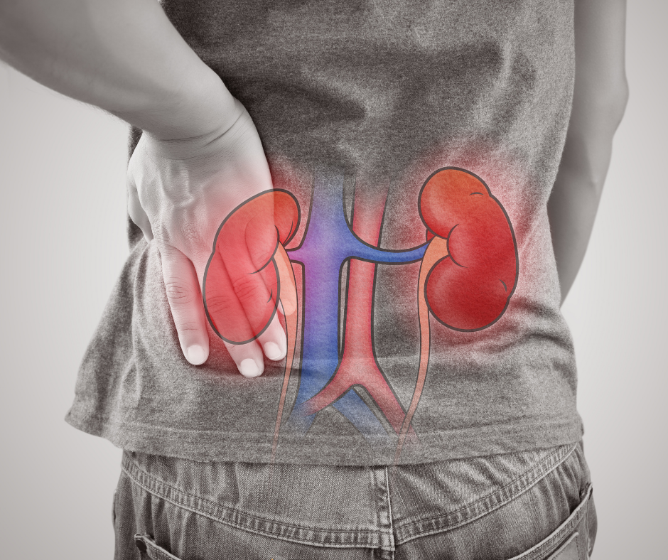
What is a kidney stone?
Kidney stones (also called renal calculi or nephrolithiasis) are hard deposits of minerals and salts that form inside your kidneys. They develop when your urine becomes concentrated, allowing minerals to crystallise and clump together. Stones vary enormously in size — from a tiny grain of sand to, in rare cases, a stone that fills the entire kidney cavity.
There are four main types:
- Calcium oxalate stones — the most common type, formed when calcium combines with oxalate in the urine
- Uric acid stones — often linked to a diet high in purines (found in red meat, shellfish, and alcohol)
- Struvite stones — typically associated with urinary tract infections
- Cystine stones — a rarer type caused by a hereditary condition
Knowing which type you have is important, because it guides both treatment and long-term prevention.
Recognising the symptoms
Not all kidney stones cause symptoms straight away. Small stones may pass unnoticed through your urine. Larger stones, however, can cause a distinctive pattern of symptoms:
- Sudden, intense pain in the back, side, or lower abdomen (known as renal colic) — often described as one of the most severe pains a person can experience
- Pain that radiates to the groin or inner thigh
- Nausea and vomiting
- Blood in the urine (haematuria), which may appear pink, red, or brown
- Frequent or painful urination
- Cloudy or foul-smelling urine
- Fever and chills — a sign that infection may be present and that urgent assessment is needed
If you are experiencing fever alongside kidney stone symptoms, please seek urgent medical attention immediately.
Your first appointment: what happens at our clinic
When you attend Urology Clinics Manchester, your consultant will begin with a thorough clinical assessment. This includes a detailed discussion of your symptoms, your medical and family history, and any previous episodes of kidney stones. The aim is to build a complete picture before recommending investigations.
You may be asked about your fluid intake, diet, and medications — all of which can influence stone formation.
Diagnosis: the investigations you may need
Accurate diagnosis is the foundation of effective treatment. Depending on your presentation, your urologist may recommend one or more of the following:
CT KUB (Computed Tomography of Kidneys, Ureters and Bladder)
This is the gold-standard imaging investigation for kidney stones. A low-dose CT scan produces detailed cross-sectional images that can detect virtually all types of stone, regardless of their composition or size. It takes only a few minutes and involves no injections or contrast dye in most cases.
Ultrasound
Ultrasound uses sound waves to produce images of the kidneys. It is radiation-free, making it particularly useful for younger patients, pregnant women, and for monitoring known stones over time. While ultrasound may not detect all small stones as reliably as CT, it is an excellent first-line tool.
X-ray (KUB)
Plain X-rays can identify calcium-containing stones that appear radio-opaque (visible on X-ray). They are often used alongside other investigations or to monitor stone position over time.
Urine and blood tests
A urine dipstick and microscopy can detect blood, infection, and crystals. Blood tests help assess kidney function (via creatinine levels) and check for underlying metabolic conditions such as hypercalcaemia (elevated calcium). If you have recurrent stones, a 24-hour urine collection may be requested to analyse your urine chemistry in detail.
Stone analysis
If you pass a stone naturally, we encourage you to collect it in a clean container and bring it to your appointment. Laboratory analysis of the stone reveals its exact composition, which is invaluable for tailoring your prevention strategy.
Treatment options: from conservative management to surgery
The right treatment depends on several factors: the size and location of the stone, your symptoms, your kidney function, and whether infection is present. At Urology Clinics Manchester, we offer the full spectrum of evidence-based treatment options.
Conservative management (watchful waiting)
Stones measuring less than 5–6 mm have a reasonable chance of passing spontaneously. Your consultant may recommend drinking plenty of fluids (ideally 2.5–3 litres per day), taking prescribed pain relief, and using a medication called an alpha-blocker (such as tamsulosin) to relax the muscles of the ureter and help the stone pass. You will be monitored closely and advised on when to seek urgent attention.
Extracorporeal Shock Wave Lithotripsy (ESWL)
ESWL is a non-invasive procedure in which focused shock waves are directed at the stone from outside the body. The waves break the stone into smaller fragments that can then pass naturally through your urine. It is typically performed as a day procedure, requires no incisions, and is particularly effective for stones in the kidney or upper ureter.
Most patients require one to three sessions. You may experience some discomfort during and after the procedure, and temporary bruising or blood in the urine is common and expected.
Ureteroscopy (URS) and laser lithotripsy
A ureteroscope is a thin, flexible telescope passed through the urethra and bladder into the ureter or kidney under general anaesthetic. Once the stone is located, a laser fibre is used to fragment it into dust or small pieces, which are either removed directly or allowed to pass naturally.
Ureteroscopy has a very high success rate and is particularly well-suited to stones in the ureter or lower kidney. Most patients go home the same day or the day after. A temporary ureteric stent may be placed to ensure drainage while any swelling settles.
Percutaneous Nephrolithotomy (PCNL)
PCNL is reserved for large or complex stones (typically greater than 2 cm) that cannot be effectively treated by ESWL or ureteroscopy. A small incision is made in the back, and a nephroscope is passed directly into the kidney to break up and remove the stone. A short hospital stay of two to three nights is usually required. Despite being the most invasive option, PCNL offers the highest stone-free rates for large stones.
Recovery and what to expect afterwards
Recovery varies depending on the treatment you have received. Most patients who undergo ESWL or ureteroscopy can return to normal activities within a few days to a week. PCNL typically requires a longer recovery of two to four weeks before returning to work.
You should expect:
- Some blood in the urine for a few days after any procedure — this is normal
- Mild discomfort or a sensation of urgency if a ureteric stent is in place
- Instructions on fluid intake, activity levels, and follow-up imaging
Your follow-up appointment will confirm that the stone has been successfully treated and allow us to discuss long-term prevention strategies.
Preventing kidney stones from coming back
Once you have had one kidney stone, your risk of developing another is significantly increased. The encouraging news is that recurrence is largely preventable with the right lifestyle adjustments and, where needed, medication.
General measures that make a real difference include:
- Drinking enough fluid throughout the day to produce at least 2–2.5 litres of urine (aim for pale, straw-coloured urine)
- Reducing dietary salt, which increases calcium in the urine
- Moderating intake of oxalate-rich foods such as spinach, nuts, and chocolate if you form calcium oxalate stones
- Limiting red meat and alcohol if you form uric acid stones
- Maintaining a healthy body weight
Depending on your stone analysis and urine biochemistry, your consultant may also recommend specific medications — for example, thiazide diuretics to reduce urinary calcium, potassium citrate to alkalinise the urine, or allopurinol to reduce uric acid levels.
Every patient is different. At Urology Clinics Manchester, we tailor prevention plans to your individual stone type and metabolic profile — because a one-size-fits-all approach simply is not good enough.
When to seek urgent help
Most kidney stones, while painful, are not immediately dangerous. However, there are situations that require same-day or emergency assessment:
- Fever or rigors (shaking chills) alongside kidney stone symptoms — this suggests infection and requires urgent treatment
- Severe, uncontrollable pain or vomiting
- A solitary kidney or known reduced kidney function
- Inability to pass urine
Book a consultation
If you are experiencing symptoms that may indicate a kidney stone, or if you have already been diagnosed and are looking for specialist guidance on treatment or prevention, our team of experienced consultant urologists is here to help.
We offer prompt appointments with access to state-of-the-art diagnostics and the full range of treatment modalities — all under one roof.


0 Comments